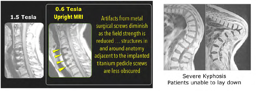
There is also a competitive advantage that relates to reducing image artifacts arising from metal implants such as surgical screws. It is well known that such artifacts get smaller as the MRI magnet’s field strength is reduced, so the anatomy adjacent to implanted hardware will be less obscured with the Upright MRI. This is particularly valuable for surgeons referring their postoperative patients for diagnostic imaging studies. In addition, image artifacts from physiological motion are similarly reduced relative to the higher field systems; note the area anterior to the spine in the kyphosis patients scanned sitting upright (since it is difficult for them to be scanned lying down).
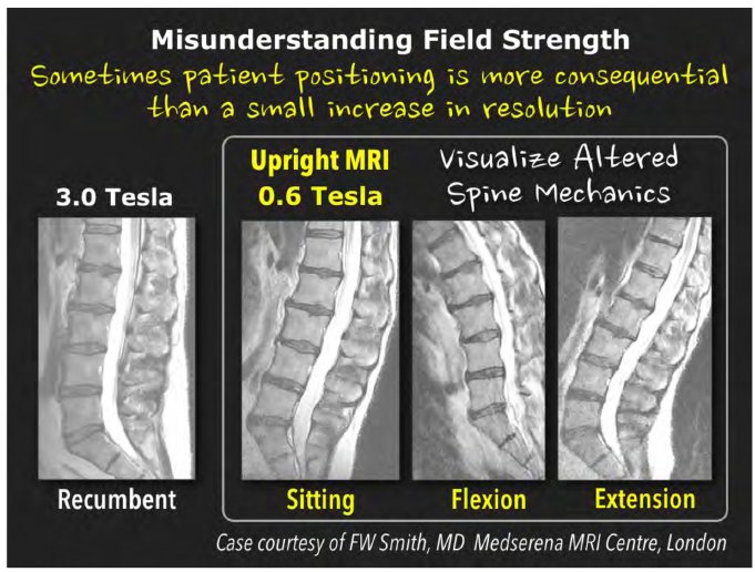
How do routine images from the 0.6 Tesla Upright MRI compare to those, say, from a high-field 3.0 Tesla IvIRI system?
There are numerous tradeoffs. For instance, as T1 NMR tissue relaxation times are known to increase with magnetic field strength, higher field magnets typically suffer from reduced T1 contrast in T1W images. On the other hand, the high-field MRI’s increased signal-to-noise means that in a fixed scan time it can obtain higher resolution images. Of course with a mid field MRI the technologist can increase the scan time to match the high resolution obtained with the higher field strength MRIs.


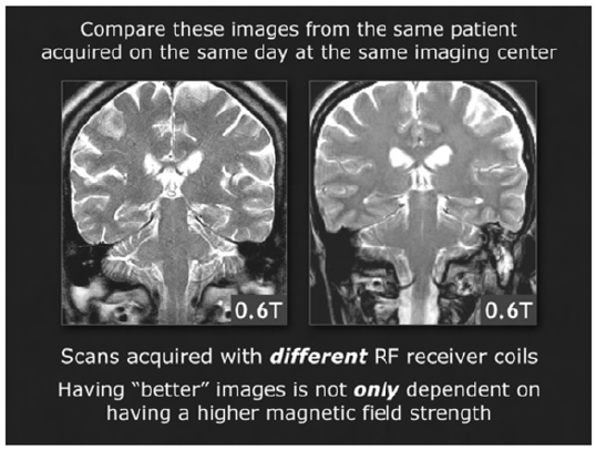
It is also important to recognize that signal-to-noise at a given field strength can be increased by incorporating innovative RF receiver coil design improvements.
The 0.6 Tesla Upright MRI’s field strength and gradient specifications are sufficient for performing specialized MRI applications.
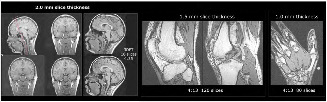
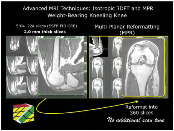
3DFT techniques are ideal for mid-field MRI systems because of their increase in signal-to-noise as well as their ability to provide thin contiguous slices.
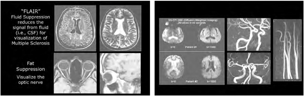
Neuroimaging applications (above) include FLAIR, fat suppression using 3-point Dixon water-fat separation techniques, diffusion-weighted imaging (DWI) and Magnetic Resonance Angiography (MRA).